

3D mammography
Tomosynthesis
Tomosynthesis produces three-dimensional cross-sectional images of the breast and detects up to 30–35 per cent more invasive tumours than conventional 2D mammography – particularly in cases of dense breast tissue.
Basics
What is tomosynthesis?
Tomosynthesis (also known as 3D mammography or digital breast tomosynthesis) is an advancement of conventional mammography and is therefore an advanced imaging technology developed specifically for the early detection of breast cancer. It represents an advancement on conventional mammography and offers significant advantages in terms of diagnostic accuracy.
In tomosynthesis, the breast is examined using X-ray-based 2D images taken from various angles. Unlike traditional mammography, which captures only single 2D images, in tomosynthesis the X-ray tube rotates around the breast and produces a series of images from multiple perspectives. These images are then combined into a 3D volume set, which enables the radiologist to ‘scroll’ through the breast tissue – much like a CT scan. This helps to avoid tissue overlaps that can obscure or mimic findings in 2D mammography.
What are the advantages of tomographic imaging?
Tomosynthesis makes it possible to visualise the breast tissue at different depths parallel to the detector surface. This filters out overlapping tissue structures, such as dense glandular tissue or connective tissue, which could mask tumours. This provides a clearer view of the tissue structure and enables a more precise diagnosis.
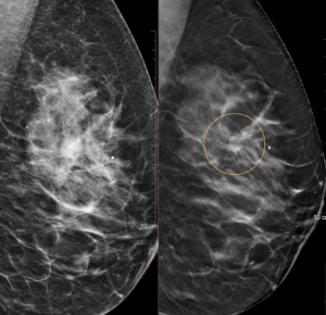
Left: 2D mammography | Right: 3D tomosynthesis
Indications
When is tomosynthesis recommended?
Tomosynthesis is particularly valuable for patients in whom 2D mammography has reached its limits.
Dense breast tissue

In cases of dense breast tissue, tomosynthesis can provide a clearer view than conventional 2D mammography. The layered images reduce tissue overlap, making it easier to detect abnormalities
Family history
Where there is an increased risk but the criteria for a breast MRI have not yet been met, tomosynthesis is a valuable intermediate step.
Investigation of unclear findings
If a finding on a mammogram cannot be clearly assessed, tomosynthesis can provide further insight thanks to its layered imaging.
Comprehensive healthcare from the age of 40
As a supplementary or alternative form of care for women who wish to undergo a more detailed examination than the statutory screening.
Reducing reorders
Tomosynthesis often produces clearer images. This helps to reduce the number of unnecessary repeat appointments due to unclear findings.
Pre-operative diagnosis
Prior to planned breast surgery, tomosynthesis provides precise information on the location and extent of a lesion.
Preparation
Please do not use deodorant, talcum powder or body lotion on your chest or underarms on the day of the examination – these can affect the image quality. The screening should not take place shortly before or during your period.

Positioning
The breast is positioned on the machine’s support plate, one side at a time, and compressed briefly. The compression is necessary to ensure clear images and takes only a few seconds.
3D images
The X-ray machine sweeps in an arc across the breast, taking a series of individual images. The images are then reconstructed and divided into layers. These layers produce a detailed 3D representation of the breast tissue.
This makes it much easier to visualise tumours and other abnormalities, even if they are obscured by dense tissue.
Findings & Report
Our radiologists will assess the images and draw up a written report. The findings will be discussed with you and/or forwarded directly to your gynaecologist.
FAQ
Questions about tomosynthesis
Digital breast tomosynthesis is not currently part of the statutory mammography screening programme and is therefore often offered as an individual healthcare service (IGeL). Whether this supplementary examination is appropriate in your case depends on your individual findings, your breast density and your personal risk profile. We would be happy to advise you based on your previous examinations.


