
Our Services
Bone Scintigraphy
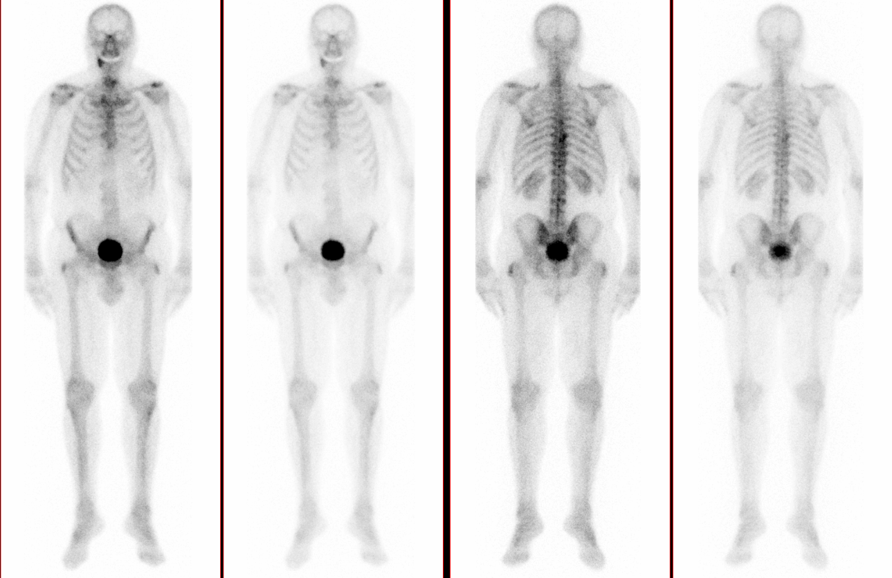
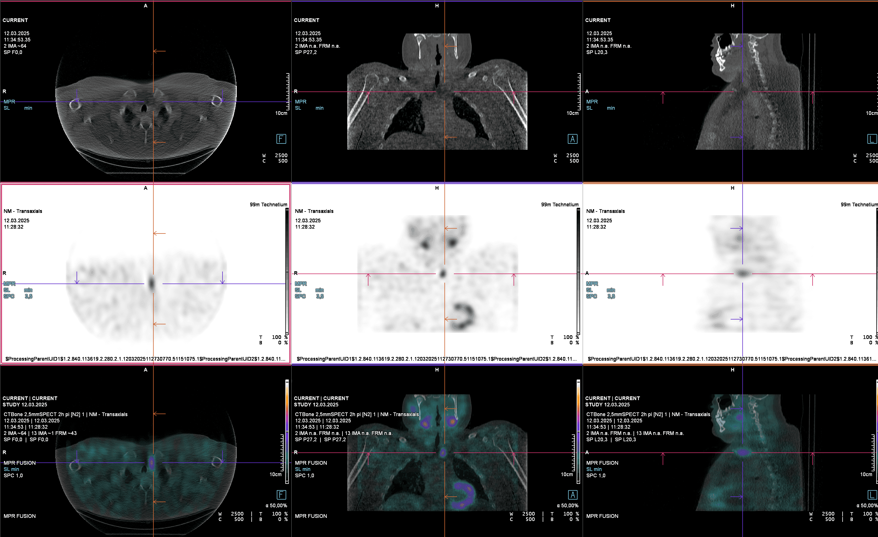
Bone Scintigraphy – Whole-Body Imaging for Bone Metastases, Fractures, and Prosthesis Loosening Bone scintigraphy (also called skeletal scintigraphy) is a nuclear medicine imaging technique that visualizes the metabolic activity of the entire skeleton.
Bone Scintigraphy
Bone Scintigraphy – Whole-Body Imaging for Bone Metastases, Fractures, and Prosthesis Loosening Bone scintigraphy (also called skeletal scintigraphy) is a nuclear medicine imaging technique that visualizes the metabolic activity of the entire skeleton.

Cardiac scintigraphy
Cardiac scintigraphy (also known as myocardial perfusion) is a non-invasive and extremely gentle examination method for imaging the blood flow in the heart muscle - in contrast to cardiac catheterization. In this way, incipient circulatory disorders (ischemia) can be detected at an early stage and known coronary heart disease can be reliably monitored.

Lung scintigraphy (V/Q scan)
Pulmonary scintigraphy can be used to painlessly detect or rule out an embolism. The examination is carried out without iodine-containing contrast medium and is therefore gentle on kidneys, hyperthyroidism and allergy sufferers.
Lung scintigraphy (V/Q scan)
Pulmonary scintigraphy can be used to painlessly detect or rule out an embolism. The examination is carried out without iodine-containing contrast medium and is therefore gentle on kidneys, hyperthyroidism and allergy sufferers.
Parathyroid scintigraphy
Parathyroid scintigraphy localizes an overactive parathyroid adenoma that increases the calcium level. Accurate detection allows precise and minor neck surgery.
Parathyroid scintigraphy
Parathyroid scintigraphy localizes an overactive parathyroid adenoma that increases the calcium level. Accurate detection allows precise and minor neck surgery.

Renal scintigraphy (MAG3 & DMSA)
Renal scintigraphy measures separately how well each kidney filters and whether urine builds up. This allows a one-sided functional weakness or obstruction of the outflow to be detected long before creatinine levels rise.
Renal scintigraphy (MAG3 & DMSA)
Renal scintigraphy measures separately how well each kidney filters and whether urine builds up. This allows a one-sided functional weakness or obstruction of the outflow to be detected long before creatinine levels rise.

Sentinel Lymph Node Scintigraphy
Sentinel Lymph Node Scintigraphy – Precision Guidance in Cancer Surgery Sentinel lymph node scintigraphy (also called sentinel node mapping) is a nuclear medicine imaging technique that identifies the first lymph node in the drainage area of a tumor. This “sentinel node” acts as the first filter where cancer cells may spread.
Somatostatin Receptor Scintigraphy (Tektrotyd®)
Targeted Imaging for Neuroendocrine Tumors Somatostatin receptor scintigraphy is a specialized nuclear medicine imaging technique used in the diagnosis of neuroendocrine tumors (NET). These tumors typically express somatostatin receptors on their surface. By injecting a small amount of radioactive somatostatin analogue (e.g., Tektrotyd®), tumors and their metastatic spread can be visualized with high precision.

Thyroid scintigraphy (+ suppression)
Thyroid scintigraphy (+ suppression) gently visualizes functional disorders and enables targeted therapy planning. It shows which thyroid nodules absorb a lot or little iodine. This allows harmless “hot” nodules to be distinguished from suspicious “cold” nodules - often saving unnecessary punctures or operations.

Ultrasound Thyroid
Thyroid ultrasound examines thyroid gland size, structure, and potential nodules or cysts.
Ultrasound Thyroid
Thyroid ultrasound examines thyroid gland size, structure, and potential nodules or cysts.
Your wellness, just a step away
Easily book your appointment online
Choose the time, location, and service you need
News & Insights

07.05.2026
Quartz Healthcare acquires the Center for Radiotherapy Dr. Meinecke in Berlin
WUPPERTAL, NRW, 07052026 – Quartz Healthcare Germany announces the integration of the Center for Radiotherapy and Radiation Oncology Dr
23.03.2026
PRESS RELEASE Quartz Healthcare Integrates Radiology Euskirchen
WUPPERTAL, NRW, 23032026 – Quartz Healthcare Germany announces the integration of Radiology Euskirchen into the group

01.10.2025
Quartz Healthcare Acquires MVZ Rheine in North Rhine-Westphalia
WUPPERTAL, NRW, 01102025 – Quartz Healthcare Germany announces the acquisition of MVZ Rheine, an established outpatient provider of radiology and nuclear medicine services in North Rhine-Westphalia

03.09.2025
radprax MVZ rebrands to Quartz: Completion of brand integration following the 2022 acquisition
Arnsberg/Wuppertal, [20 August 2025] – The radprax companies are rebranding to Quartz Westfalen MVZ GmbH (formerly radprax MVZ Westfalen GmbH) with headquarters in Arnsberg, and Quartz Nordrhein MVZ GmbH (formerly radprax MVZ Nordrhein GmbH) based in Wuppertal

17.07.2025
PRESS RELEASE: Quartz Healthcare Germany Acquires Radiologie Mansfelder Land
Quartz Healthcare Germany Acquires Radiologie Mansfelder Land – Expanding Presence in Saxony-Anhalt Wuppertal, NRW, July 17, 2025 – *Quartz Healthcare Germany announces the acquisition of Radiologie Mansfelder Land, a leading outpatient radiology services provider in Saxony-Anhalt

15.12.2024
Herniated Disc: How Radiology Can Help
Herniated Disc: How Radiology Can Help A herniated disc might sound like an acute and sudden issue, but it is often the result of a gradual wear-and-tear process
12.12.2024
Parkinson’s Disease: Diagnosis Through Radiology
Parkinson’s disease is one of the most frequently diagnosed neurological disorders, particularly in individuals over the age of 60
12.12.2024
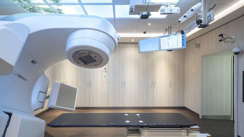
Radiation Therapy: How It Works and Possible Side Effects Explained
Radiation Therapy: How It Works and Possible Side Effects Explained Radiation therapy (radiotherapy) is one of the three main pillars of modern cancer treatment, alongside chemotherapy and surgical tumor removal